Introduction
The human eye is one of the most fascinating and complex organs in the body. Every blink, every glance, and every color perceived depends on a delicate structure that works in perfect harmony to capture and process light.

From the outermost layer that protects the eyeball to the intricate network of nerves that transmit visual information to the brain, each part of the eye has a distinct and vital role. The process of seeing is not merely about the eye—it involves the coordination between the and the brain, transforming light into the beautiful, colorful world that surrounds us.
Eye Anatomy Overview
The functions much like a sophisticated camera.
The eye can be divided into several regions:
- The front portion, which includes the cornea, anterior chamber, iris, pupil, and lens
- The middle layer, known as the uvea, containing blood vessels and muscles
- The back portion, which includes the retina, macula, optic nerve, and vitreous body
Together, these elements form a highly coordinated visual system.
Parts of the Eye Outside the Eyeball
The human eye is protected by a strong, bony structure called the orbit, commonly referred to as the eye socket. The orbit houses the eyeball and cushions it with layers of fat and connective tissue, ensuring it remains stable yet flexible.
Surrounding the are six extraocular muscles, responsible for its movement in every direction. These muscles enable the eye to move up, down, sideways, and diagonally. Each muscle works in coordination with its counterpart to ensure smooth, precise movements.
Blinking spreads tears evenly across the surface of the eye, keeping it moist and clean.
The Surface of the Eye

This thin tissue acts as a protective barrier, shielding the from dust, microorganisms, and other irritants.
The eye’s surface is kept moist and nourished by a remarkable system known as the tear film. The tear film is made up of three layers:
- The mucous layer – produced by the conjunctiva, it helps tears spread evenly.
- The aqueous layer – formed by the lacrimal gland, located beneath the outer edge of the eyebrow; this watery layer hydrates and nourishes the cornea.
- The lipid (oil) layer – created by the meibomian glands, which prevents tears from evaporating too quickly.
This is why crying often leads to a runny nose.
Maintaining a healthy tear film is essential for comfort and clear vision, as even minor dryness can cause irritation, redness, or blurred sight.
The Front of the Eye
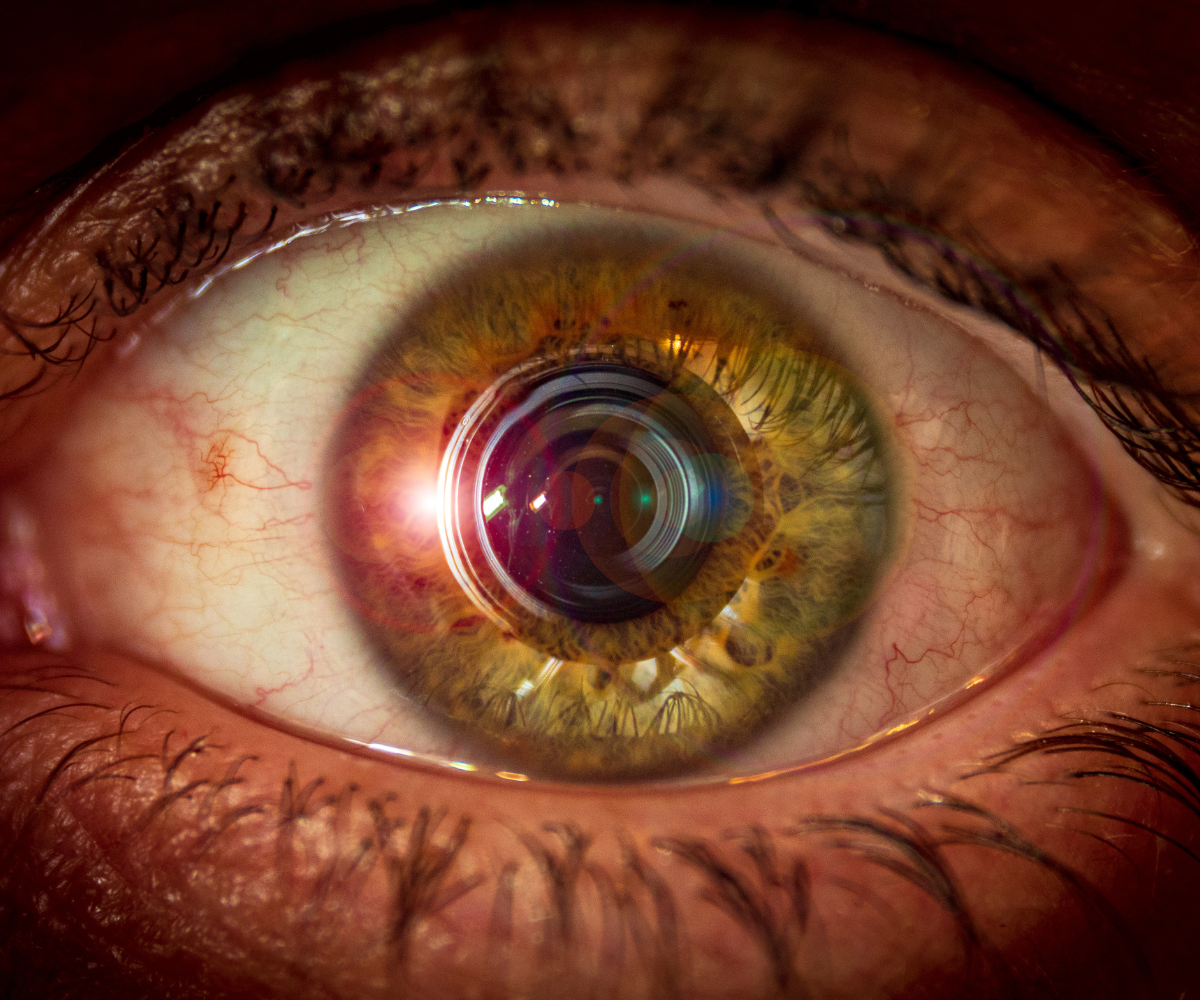
The front part of the eye is the first to interact with light. The cornea—a transparent, dome-shaped layer—acts as the eye’s window. It bends (refracts) incoming light toward the center, providing about 70% of the eye’s total focusing power.
Behind the anterior chamber sits the iris, the colored part of the that surrounds the pupil, the dark circular opening in its center. The iris contains small muscles that control pupil size—widening (dilating) in dim light and narrowing (constricting) in bright conditions. This automatic adjustment helps regulate how much light enters the eye.
Just behind the pupil is the lens, a transparent, flexible structure that fine-tunes focus. The lens changes shape through the action of small fibers called zonules, connected to a circular muscle known as the ciliary body. This process, known as accommodation, allows the eye to switch focus between distant and nearby objects.https://www.youtube.com/watch?v=TY1giZgddAs
The Middle Layer: The Uvea
Beneath the sclera lies the uvea, the middle layer of the eye. It consists of three key components:

- The iris, which regulates light intake.
- The ciliary body, which produces aqueous humor and helps focus the lens.
- The choroid, a dense network of blood vessels that nourishes the retina.
The choroid provides oxygen and nutrients to the outer layers of the retina.
The Back of the Eye
This material helps the eye maintain its spherical shape and provides support for the retina.
At the very back lies the retina, a delicate tissue responsible for capturing light and converting it into electrical impulses. Within the retina are specialized cells known as photoreceptors, of which there are two types:
- Rods, which detect light intensity and enable night vision.
- Cones, which perceive color and detail, essential for daytime vision and reading.
The macula, a small area in the retina, provides the sharp central vision needed for recognizing faces and reading. The peripheral retina helps detect motion and gives us side vision.

The optic nerve, made up of over a million nerve fibers, carries these signals to the visual cortex, where the brain interprets them as images.
This intricate connection between the eye and brain allows humans to perceive depth, color, motion, and shape in extraordinary detail.