Eye Anatomy: Parts of the Eye and How Vision Works
Understanding eye anatomy is essential for appreciating how vision works and how the eye interacts with the brain to create the sense of sight. The human eye is a complex organ composed of multiple layers, fluids, and specialized tissues that work together to focus light, detect images, and transmit information to the brain. This guide provides a detailed look at the parts of the eye, their functions, and the mechanisms behind human vision.
Parts of the Eye Outside the Eyeball
The eye sits within a protective bony cavity called the orbit, which shields it from injury. Surrounding the eyeball are six extraocular muscles that allow precise movement in multiple directions. These muscles are anchored to the sclera, the tough white outer layer of the eyeball.
Extraocular Muscles
- Superior rectus: moves the eye upward.
- Inferior rectus: moves the eye downward.
- Medial rectus: moves the eye inward, toward the nose.
- Lateral rectus: moves the eye outward, away from the nose.
- Superior oblique: rotates the eye downward and outward.
- Inferior oblique: rotates the eye upward and outward.
These muscles allow smooth coordination for tracking moving objects, reading, and performing daily tasks.
Eyelids and Conjunctiva
The inner surface of the eyelids and the outer surface of the eyeball are covered with a clear protective membrane called the conjunctiva. This thin layer protects the eye from dust, microorganisms, and other harmful particles while contributing to tear production.
The Surface of the Eye and Tear Film
The tear film is a three-layered fluid that maintains lubrication and protects the cornea from infection:
- Mucous layer: Produced by goblet cells in the conjunctiva; helps the tears stick to the eye.
- Aqueous layer: Produced by the lacrimal glands located under the outer edges of the eyebrows; provides moisture and nutrients.
- Oil layer: Produced by the meibomian glands along the eyelid margins; prevents evaporation of tears.
Tears drain through the nasolacrimal duct, helping keep the eye surface clean and moist. Proper tear production is crucial for comfort and clear vision.
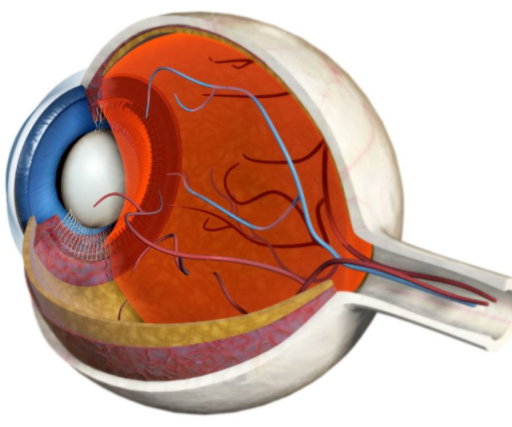
The Front of the Eye
The cornea is the transparent, dome-shaped front layer of the eye that bends light to help focus images on the retina. It is avascular, meaning it does not contain blood vessels, and relies on tears for oxygen and nutrients.
Anterior Chamber and Aqueous Humor
Behind the cornea lies the anterior chamber, filled with aqueous humor, a clear fluid that maintains intraocular pressure, provides nutrients, and removes waste. The fluid drains through the trabecular meshwork in the drainage angle to maintain proper eye pressure.
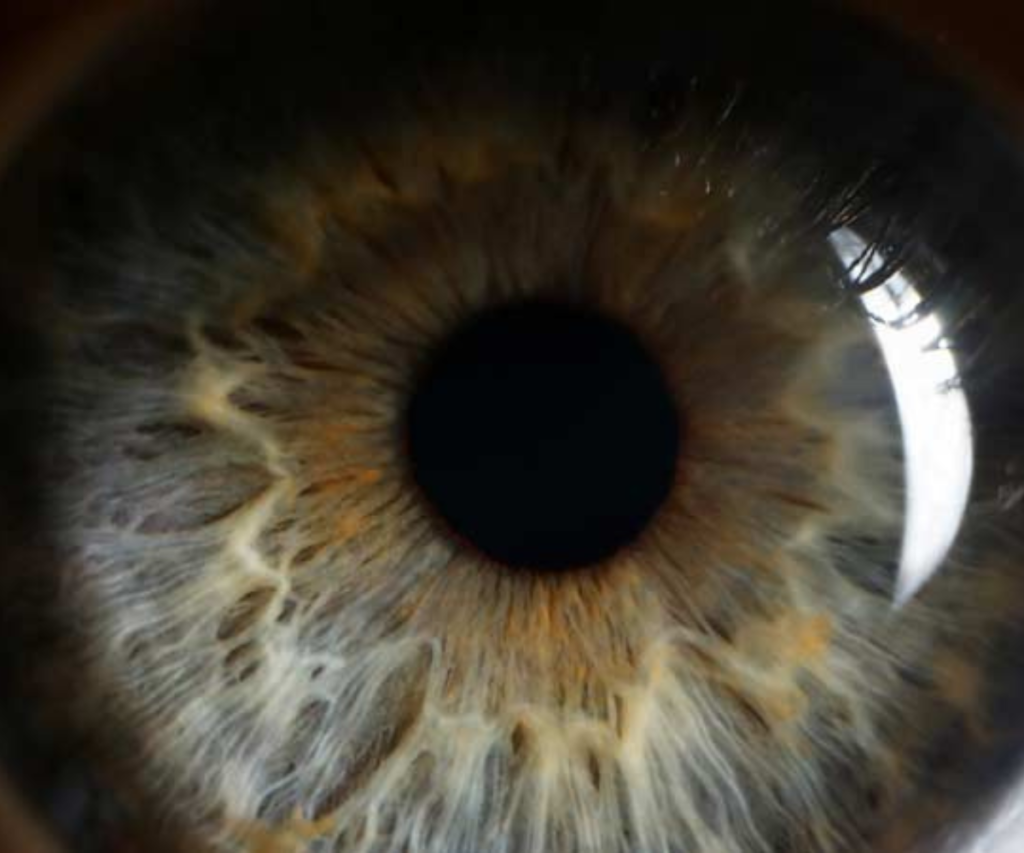
Iris and Pupil
The iris, the colored part of the eye, controls the size of the pupil, the central opening through which light enters. Muscles in the iris dilate or constrict the pupil to regulate light intake, enabling vision under various lighting conditions.
Lens
The lens lies directly behind the pupil and fine-tunes focus by changing shape, a process called accommodation. Fibers known as zonules hold the lens in place, allowing it to adjust for near and distant objects. During cataract surgery, the natural lens may be replaced with an intraocular lens that fits inside the lens capsule.
Focus Contribution: The cornea provides approximately 70% of the eye’s focusing power, while the lens contributes 30%. Both work together to create a sharp image on the retina.
The Back of the Eye
Vitreous Humor and Vitreous Cavity
The vitreous cavity occupies the space between the lens and the retina and is filled with vitreous humor, a jelly-like substance that maintains the eye’s shape and supports the retina.
Retina and Macula
The retina is the light-sensitive layer lining the back of the eye. It contains photoreceptor cells that convert light into electrical signals:
- Rods: Detect black and white light, enabling night vision.
- Cones: Detect color and detail, allowing central vision.
The macula, a specialized area in the center of the retina, is responsible for high-resolution vision and tasks like reading, recognizing faces, and seeing fine details.
Optic Nerve and Visual Cortex
Electrical signals from photoreceptors travel via the optic nerve to the visual cortex in the brain, where they are interpreted as images. Each optic nerve contains over a million fibers that transmit information rapidly, enabling real-time vision.
Supporting Structures of the Eye

Sclera and Choroid
The sclera is a dense, white protective layer covering most of the eyeball. Beneath it lies the choroid, a layer rich in blood vessels that provides oxygen and nutrients to the retina.https://www.youtube.com/watch?v=TY1giZgddAs
Ciliary Body and Zonules
The ciliary body produces aqueous humor and contains muscles that adjust lens shape. Zonules connect the ciliary body to the lens capsule, enabling accommodation for clear near and far vision.
Optic Disc
The optic disc is where the optic nerve exits the eye. It contains no photoreceptors, creating a natural blind spot in each eye.
How the Eye Works: From Light to Vision
- Light enters through the cornea.
- The pupil adjusts to control the amount of light.
- The lens fine-tunes focus on the retina.
- The retina’s photoreceptors convert light into electrical impulses.
- Impulses travel through the optic nerve to the brain.
- The visual cortex interprets these signals as images.
This intricate process allows humans to perceive the world in color, depth, and detail.
Common Eye Conditions Related to Anatomy
- Cataracts: Clouding of the lens, affecting vision clarity.
- Glaucoma: Increased intraocular pressure damaging the optic nerve.
- Macular Degeneration: Degeneration of the macula, reducing central vision.
- Retinal Detachment: Separation of the retina from underlying tissues, a medical emergency.
- Dry Eye Syndrome: Insufficient tear production, affecting lubrication and comfort.
Understanding eye anatomy is essential for diagnosing and managing these conditions effectively.
Eye Care Tips for Healthy Vision
- Regular eye exams.
- Protect eyes from UV light using sunglasses.
- Maintain proper screen distance and lighting.
- Stay hydrated to support tear production.
- Eat a diet rich in vitamin A, C, and omega-3 fatty acids.
- Avoid smoking, which increases risk of macular degeneration.
- Use protective eyewear during sports or hazardous activities.
Conclusion
The human eye is a remarkable organ, integrating complex structures to process light into meaningful images. From the cornea and lens focusing light, to the retina converting light into electrical signals, and the optic nerve transmitting vision to the brain, every part has a vital role. Understanding eye anatomy can enhance eye care, promote early detection of diseases, and improve overall visual health.